


 Foundational characteristics of cancer include proliferation, angiogenesis, migration, evasion of apoptosis, and cellular immortality. Find key markers for these cellular processes and antibodies to detect them.
 The SUMOplot™ Analysis Program predicts and scores sumoylation sites in your protein. SUMOylation is a post-translational modification involved in various cellular processes, such as nuclear-cytosolic transport, transcriptional regulation, apoptosis, protein stability, response to stress, and progression through the cell cycle.
 The Autophagy Receptor Motif Plotter predicts and scores autophagy receptor binding sites in your protein. Identifying proteins connected to this pathway is critical to understanding the role of autophagy in physiological as well as pathological processes such as development, differentiation, neurodegenerative diseases, stress, infection, and cancer.
LRIG3 Antibody (N-Term)
Purified Rabbit Polyclonal Antibody (Pab)
- SPECIFICATION
- CITATIONS
- PROTOCOLS
- BACKGROUND

Application
| WB, E |
|---|---|
| Primary Accession | Q6UXM1 |
| Reactivity | Human |
| Host | Rabbit |
| Clonality | polyclonal |
| Isotype | Rabbit IgG |
| Calculated MW | 123434 Da |
| Gene ID | 121227 |
|---|---|
| Other Names | Leucine-rich repeats and immunoglobulin-like domains protein 3, LIG-3, LRIG3, LIG3 |
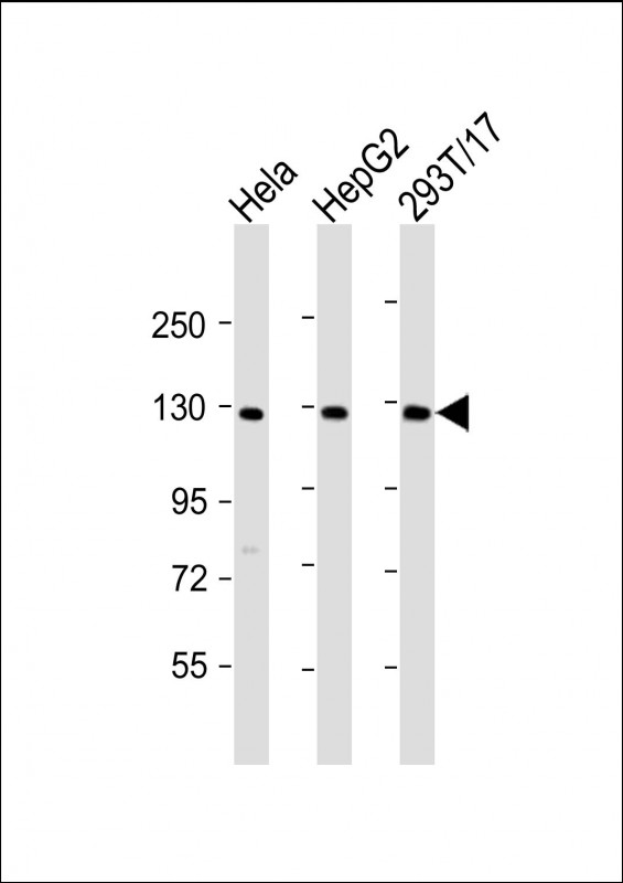
| Target/Specificity | This LRIG3 antibody is generated from a rabbit immunized with a KLH conjugated synthetic peptide between 23-54 amino acids from human LRIG3. |
| Dilution | WB~~1:2000 E~~Use at an assay dependent concentration. |
| Format | Purified polyclonal antibody supplied in PBS with 0.09% (W/V) sodium azide. This antibody is purified through a protein A column, followed by peptide affinity purification. |
| Storage | Maintain refrigerated at 2-8°C for up to 2 weeks. For long term storage store at -20°C in small aliquots to prevent freeze-thaw cycles. |
| Precautions | LRIG3 Antibody (N-Term) is for research use only and not for use in diagnostic or therapeutic procedures. |
| Name | LRIG3 |
|---|---|
| Synonyms | LIG3 |
| Function | May play a role in craniofacial and inner ear morphogenesis during embryonic development. May act within the otic vesicle epithelium to control formation of the lateral semicircular canal in the inner ear, possibly by restricting the expression of NTN1 (By similarity). |
| Cellular Location | Cell membrane; Single-pass type I membrane protein. Cytoplasmic vesicle membrane; Single-pass type I membrane protein Note=Detected in cytoplasmic vesicles when coexpressed with ERBB4 |
| Tissue Location | Widely expressed.. |

Thousands of laboratories across the world have published research that depended on the performance of antibodies from Abcepta to advance their research. Check out links to articles that cite our products in major peer-reviewed journals, organized by research category.
info@abcepta.com, and receive a free "I Love Antibodies" mug.

Provided below are standard protocols that you may find useful for product applications.
Background
May play a role in craniofacial and inner ear morphogenesis during embryonic development. May act within the otic vesicle epithelium to control formation of the lateral semicircular canal in the inner ear, possibly by restricting the expression of NTN1 (By similarity).
References
Guo D.,et al.Genomics 84:157-165(2004).
Clark H.F.,et al.Genome Res. 13:2265-2270(2003).
Ota T.,et al.Nat. Genet. 36:40-45(2004).
If you have used an Abcepta product and would like to share how it has performed, please click on the "Submit Review" button and provide the requested information. Our staff will examine and post your review and contact you if needed.
If you have any additional inquiries please email technical services at tech@abcepta.com.
Ordering Information
Shipping Information
